“I wanted to save lives”
When Muyinatu A. Lediju Bell ’06 won the National Science Foundation’s Alan T. Waterman Award, the country’s top honor for early-career researchers, she sat on a panel with her two fellow awardees, surrounded by the academic luminaries on the National Science Board. The atmosphere was formal, even weighty, but when asked by a board member how she became interested in science, Bell didn’t hesitate:
“Watching the scientists on Sesame Street mix colorful liquid in test tubes.”
Decades later, her scientific interests range across many disciplines, yet Bell’s focus is singular: She wants to save millions of lives around the world by providing better tools for early detection of diseases.
The Waterman Award recognized Bell for pioneering innovations in ultrasound and photoacoustic imaging that have led to new techniques and improved the quality of medical images, especially for people with darker skin or larger bodies. She is working to ensure that those innovations eventually become accessible to everyone.
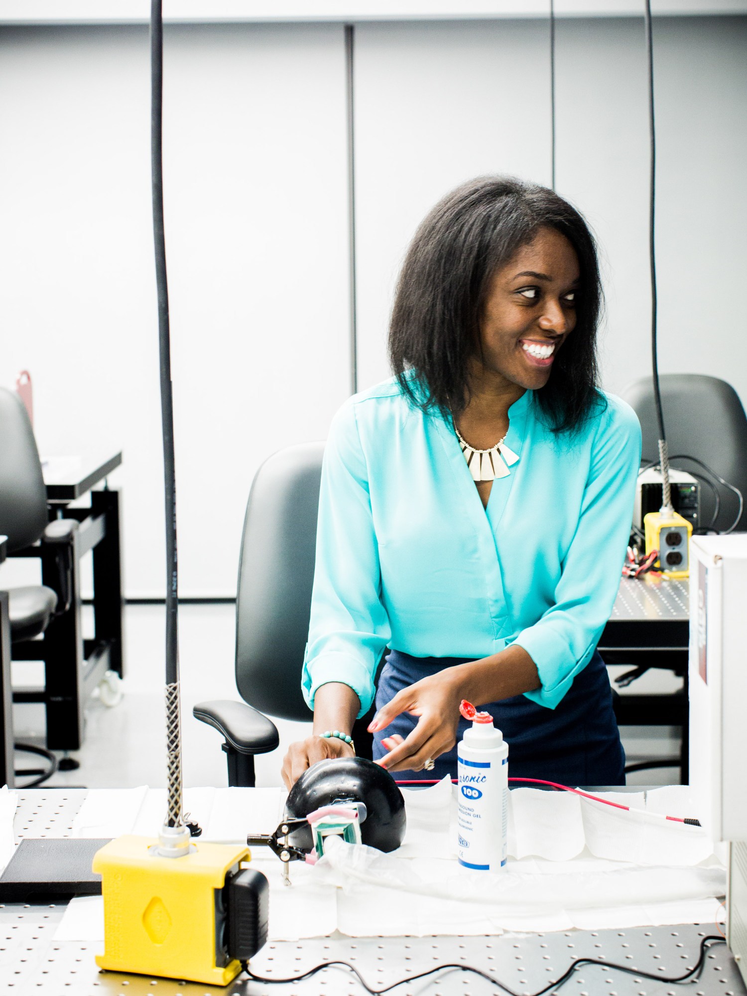
Bell, who goes by “Bisi,” is the John C. Malone Associate Professor of Electrical and Computer Engineering at Johns Hopkins University, where she founded and directs the Photoacoustic & Ultrasonic Systems Engineering (PULSE) Lab and has appointments in the departments of biomedical engineering, computer science, and oncology. On a warm day this past June she sat in her office, meeting with one of her dozen or so graduate and postdoctoral students and checking in on the student’s ongoing work.
“We work not only in optics but acoustics, robotics, signal processing, deep learning, and AI,” she said, walking through one of the four labs and centers she oversees. “We do a little bit of physics and some hands-on building and mechanical engineering.”
Bell’s goal of improving medical imaging wasn’t one she had in mind when she arrived at MIT as an undergrad, but it’s now underpinned and motivated by personal loss.
Her mother, Jane Rivers, grew up in McClellanville, South Carolina, before eventually moving to Brooklyn, where Bell was born. Rivers, who was the first in her family to go to college, spotted her daughter’s aptitude for science and math, cultivating it from an early age.
“She made it a point, very early on, to buy educational books and videos that I found very intriguing and take me on trips to the public library, where I got to dig deeper into science,” Bell says.
In grade school, Bell would do homework assigned to her brother—who was two grades ahead. “I wanted to work out his multiplication tables,” she remembers. “I always found it to be fun.” A few years later, she would tackle the math sections of her brother’s SAT prep books. “Even doing the math problems in there was fun, and it augmented what I was doing in school,” she says.
At 14, Bell enrolled in the highly selective Brooklyn Technical High School, where she divided her time between academics and a newfound interest in running; she was, as she puts it, “competitive in track and competitive in school.” During her sophomore year, she participated in a program designed to introduce girls to engineering—and was hooked. “I fell in love with engineering through that program,” she says.
Once she’d set her sights on becoming an engineer, Bell says, her brother, Abdul-Rahman Lediju (who is now an attorney), directed her attention to the Institute. “He was the one who told me about MIT,” she says, remembering her excitement. “I thought: ‘I want to cure cancer and AIDS—and I’ll get to do math there!’”
At 18, Bell already knew she wanted to focus on biomedical engineering, but at the time it was only offered as a minor, so she majored in mechanical engineering. “Just looking at the trajectory of the curriculum, I knew it was a way to get a foundation in how to make stuff in general,” she says, remembering the sequence of courses, from Mechanics and Materials to Measurement and Instrumentation. “I knew I could learn biomedical engineering later. For me it was the perfect setup.”
In her junior year, while Bell was making her way through that curriculum, her mother died of breast cancer. The loss solidified her interest in cancer research and clarified her academic path forward. “I wanted to use everything I learned at MIT, and I wanted to save lives,” she says. “So I moved to early detection and ultrasound as the best possible tool, in terms of safety, portability, and cost-efficiency.”
Bell found the right next step at Duke University in the lab of biomedical engineer Gregg Trahey, whose work focuses on developing new ultrasound technologies. She knew his lab was the ideal fit even though she jokes, “I probably scared him with how direct and focused I was.”
Bell remembers her excitement at the prospect of going to MIT. “I thought: ‘I want to cure cancer and AIDS—and I’ll get to do math there!’”
During her first year in Trahey’s lab, Bell investigated what’s known as acoustic clutter—random noises or artifacts that are recorded and translated into ultrasound images and can interfere with their clarity and usefulness. “It makes it difficult to identify structures of interest,” she explains.
But a solution soon presented itself. Bell realized that when the motion of an ultrasound probe caused the abdominal wall to move while, say, the bladder was being imaged, some of these acoustic artifacts “moved” in the image too. Analyzing that movement led to ways of filtering out that clutter, resulting in clearer ultrasonic images with better contrast-to-noise ratio, one of the main metrics of ultrasound image quality. “What’s left behind is the structure itself,” she says.
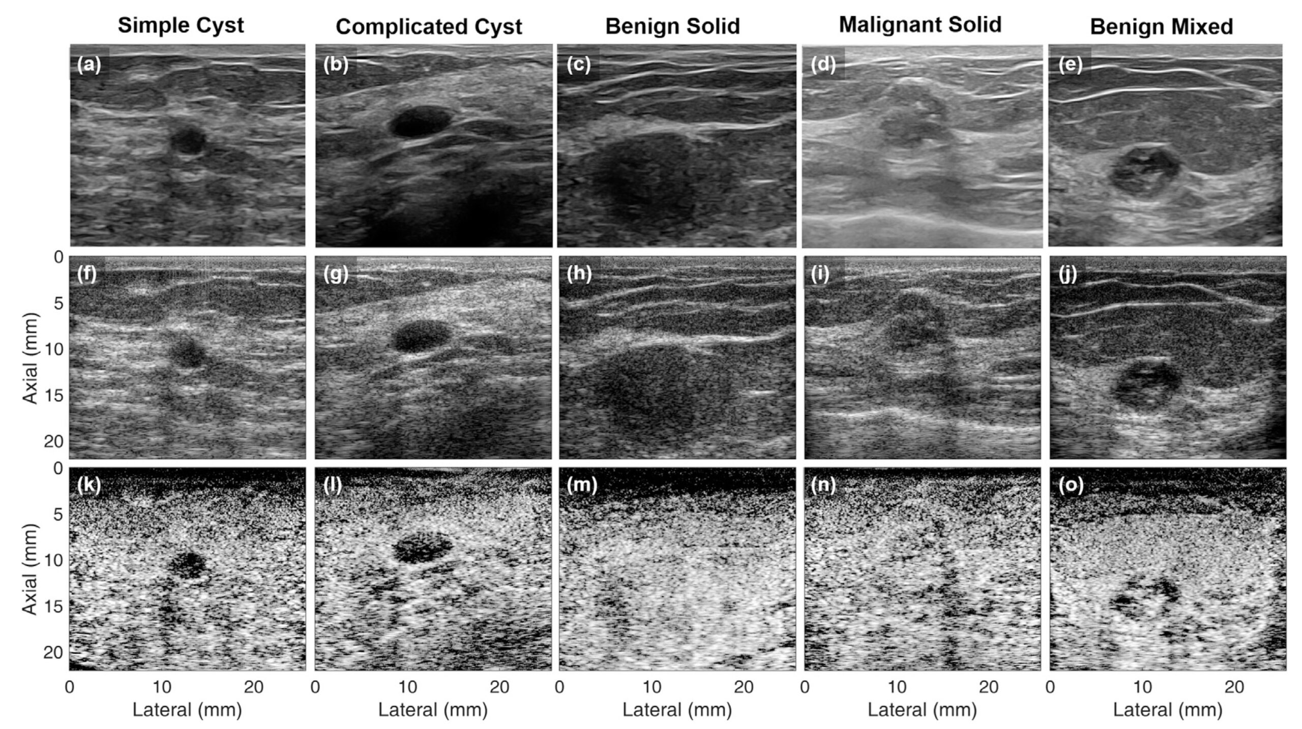
One of Bell’s pioneering discoveries came during her final years at Duke, where she developed a technique known as short-lag spatial coherence beamforming. In ultrasonography, sound waves are transmitted through the body, and echoes that bounce off internal organs are used to form images of them. These images, traditionally, are created through a process known as “delay and sum” beamforming—a signal-processing algorithm that converts the acoustic echoes captured or received by an ultrasound transducer into an image that is displayed.
Between ultrasound sensors placed on the body and the target body structure that’s being imaged, signals are being recorded at varying times. “Therefore,” Bell explained at her Waterman Award lecture in August, “we apply system time delays to align all the signals that came from the same location, and then we sum across channels to get one large high-amplitude signal associated with that location.” This process of amplitude-based delay-and-sum beamforming underlies ultrasound systems used worldwide.
But fat tissue and dense tissue can cause sound waves to bounce off an image target, distorting the final image because of acoustic scattering. This scattering is clutter that’s captured as a “random recording.” And when the random recording gets added into the summed signal that’s ultimately converted into the ultrasound image, it can obscure the target signal, potentially confounding diagnoses or treatment decisions. As a result, the traditional process isn’t as effective for larger patients and those with dense breast tissue.
Bell figured out that the signals reflecting from the targeted area of the body have a high spatial coherence, meaning that they are similar to one another. In contrast, the signals correlated with the “random recording” are less similar—they have a low spatial coherence. This discovery is the basis of the coherence-based beamforming method she invented, initially for ultrasound imaging and later for other diagnostic interventions.
By directly displaying these differences, Bell’s coherence-based approach filters out clutter and makes it possible to capture a clearer image of the target. She demonstrated the benefits in breast imaging and says that recent work has shown that the technique can be applied to ultrasounds of the liver, fetal structures, small vessels or arterioles that surround major arteries, and the heart.
Doctors will often order a follow-up ultrasound if something abnormal is found in a regular screening mammogram. In breast ultrasound, the traditional amplitude-based approach produces images in which solid masses and surrounding fluid are similarly dark. But Bell’s coherence-based technique produces a clearer image by distinguishing between the spatial coherence of fluid masses and solid masses—fluid has a lower spatial coherence than the surrounding tissue, while solid masses have a similar spatial coherence to surrounding tissue.
“This coherence-based technology is particularly promising for women with dense breasts, who tend to generate more acoustic clutter and also represent a patient population who are underserved with respect to noninvasive methods to detect breast cancer,” Bell explains.
The goal, she says, is to reduce the number of false positives and the need for biopsies or other invasive interventions, which often delay accurate diagnoses. “I would love for it to be in every clinic around the world,” she says of the technique.
After finishing her PhD at Duke in 2012, Bell went to Johns Hopkins as a postdoc in computer science and began building on the work she did in graduate school. At first she focused primarily on improving ultrasound and other diagnostics, mainly for breast cancer. She also began to apply her coherence-based beamforming technique to photoacoustics, an emerging technique for disease diagnosis that combines optical imaging with the high penetration and spatial resolution of ultrasound. As she describes it, light is transmitted to a target, which absorbs the light, undergoes thermal expansion, and generates a sound wave.
“It’s kind of like lightning and thunder, just on a smaller scale,” says Bell, who joined the Johns Hopkins faculty in 2016. “It’s the conversion of optical energy (lightning) to acoustic (thunder). We convert the optical to the acoustic.”
Bell’s short-lag spatial coherence (SLSC) beamformer technique, when integrated into photoacoustics, addresses a challenge that is common to all optics-based technologies: “poor optical penetration depth in the presence of highly melanated skin.”
Darker skin acts as an optical absorber, she explains, and the resulting photoacoustic effect propagates additional unwanted sound waves that generate acoustic clutter, which in turn obscures the signals of interest in the body.
“When we instead take the SLSC approach to imaging, we can see that regardless of skin tone, we can see the structures within the body more clearly,” Bell explains. “So while the amplitude-based approach leads to an inherent skin-tone bias, the coherence-based approach could address this bias by reducing clutter, which is particularly beneficial for patients with darker skin tones.”
The technique also gets around the scattering caused by fat tissue, leading to clearer photoacoustic imaging for people who are overweight or obese.
For Bell, considering the needs of all types of patients leads to better outcomes for a greater breadth of the population. The same is true, she explained in her Waterman lecture, of making sure that diverse types of people work in medical research: “It’s this diversity of thought that allows us to ask a diversity of questions, and it’s this diversity that really determines the type of science that’s being done.”
In addition to improving imaging techniques for traditionally marginalized groups of patients, Bell pioneered integration of photoacoustic imaging into the da Vinci surgical robot. This makes it easier for doctors to visualize such things as blood vessels, contrast agents, or metal implants in real time during surgery, making complex surgical procedures less risky. She was also the first to apply deep learning to ultrasound and photoacoustic image formation and, with her group, recently developed a way to use AI to detect covid in ultrasound images of patients’ lungs.
Bell’s 44-page CV notes her eight patents and the nearly $14 million in research funding she’s received so far, as well as an arm’s-length list of awards, including a 2016 position on MIT Technology Review’s annual list of 35 innovators under 35.
And she continues to push the boundaries of her fields, all the while trying to make diagnosing diseases more equitable and accessible.
“I hope one day,” she says, “we’ll all have ultrasounds at home and we can diagnose ourselves.”